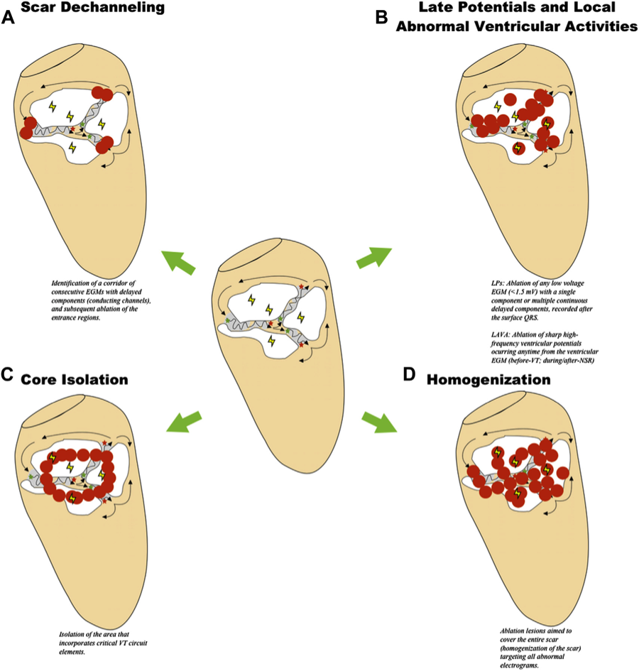
Areas between channels often have electrical activity that give rise to VT substrates (thunderbolts). Elimination of scar-related potentials is a relevant ablation goal and is the basis of different substrate-based ablation strategies.
Scar Dechanneling
Identification of a corridor of consecutive electromyograms (EGM) with delayed components (conducting channels) and subsequent ablation of the entrance regions.
(See above: A)
Late Potentials and Local Abnormal Ventricular Activities (LAVA)
Late Potentials (LPs): Ablation of any low voltage EGM (<1.5 mV) with a single component or multiple continuous delayed components recorded after the surface QRS.
Late Potentials and Local Abnormal Ventricular Activities (LAVA): Ablation of sharp high-frequency ventricular potentials occurring anytime from the ventricular EGM (before-VT; during/after normal sinus rhythm (NSR).
(See above: B)
Learn More
Core Isolation
Isolation of the area that incorporates VT circuit elements.
(See above: C)
Homogenization
Ablation lesions aimed to cover the entire scar (homogenization of the scar), thereby targeting all abnormal electrograms.
(See above: D)
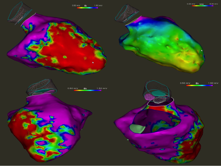
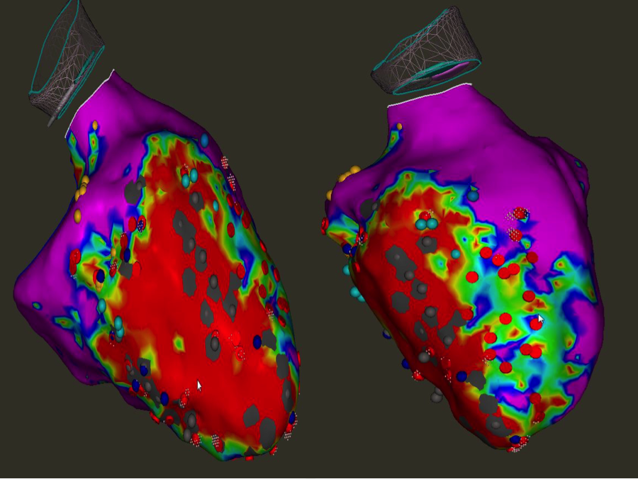
Electroanatomic voltage map of the left ventricle
Top left, bottom left and right illustrate the bipolar map in septal anterior and inferior views of the scar in the left ventricle. Top right depicts the unipolar voltage map of the left ventricle suggesting large transmural component of the scar. Septal and anterior views of the left ventricle showing radiofrequency ablation (RFA) lesions to perform substrate modification of the left ventricle (LV) septal scar. Red dots: RF lesions, gray tags denote areas or unexcitable scar.


